
Echocardiography
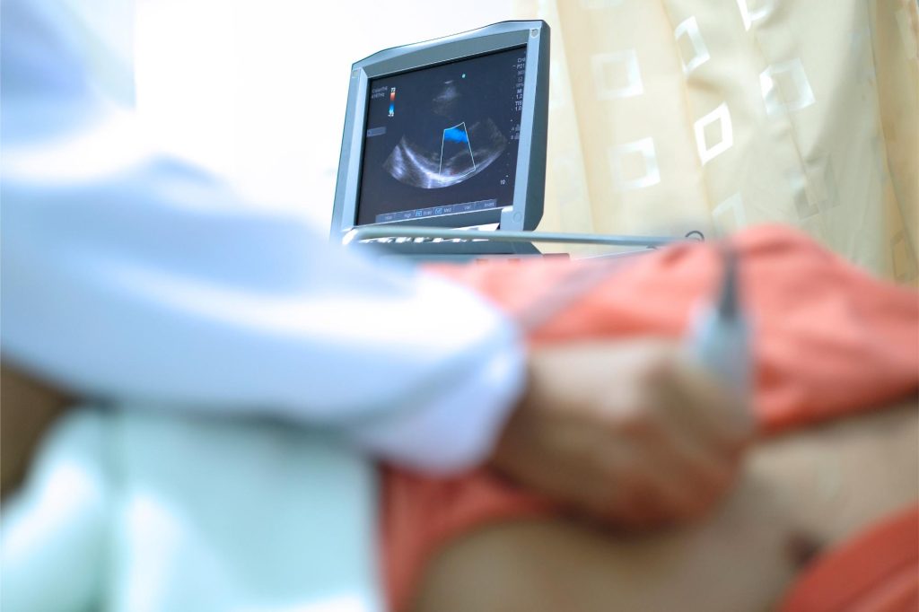
Echocardiography is a non-invasive imaging test that uses ultrasound waves to create real-time pictures of the heart. It is used to evaluate heart structures (muscles, valves), pumping function, and blood flow, to detect abnormalities. There are two main types of echocardiography: Transthoracic Echocardiography and Transesophageal Echocardiography.
Transthoracic Echocardiography
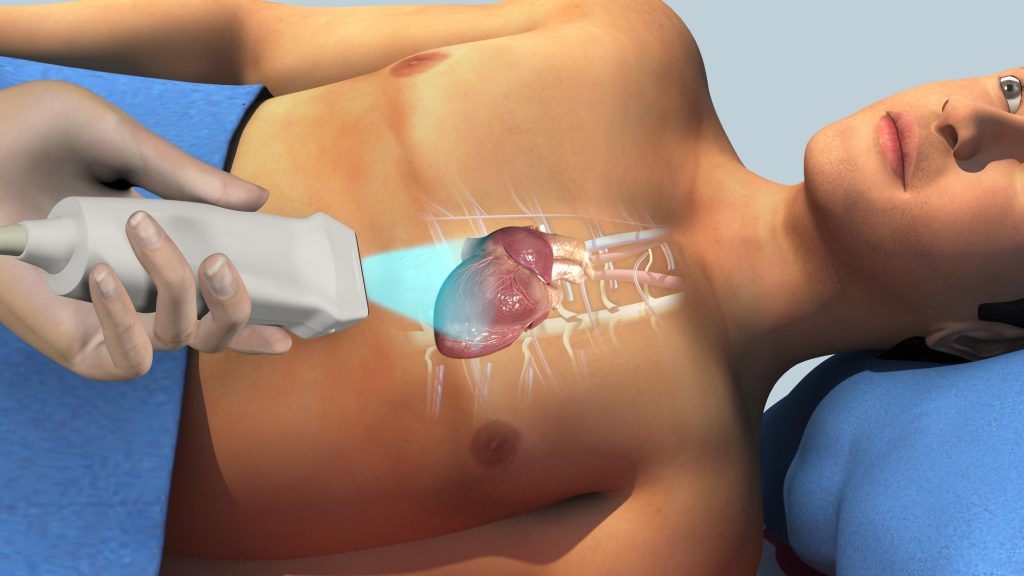
This is the most common type of cardiac ultrasound. A handheld probe (transducer) is placed on the chest wall to send and receive ultrasound waves. Transthoracic echocardiography is usually used as the first-line test for most cardiac evaluation. Its main advantages are that it is non-invasive, painless, and quick (20-30 mins). It is done without the need for sedation. However, its image quality may be limited in individuals who are obese, with thick chest wall or lung disease.
What can Echocardiography show?
- Heart chambers sizes and muscle thickness
- Heart’s pumping function
- Heart valves structure and movement
- Fluid around the heart (pericardial effusion)
- Congenital heart defects
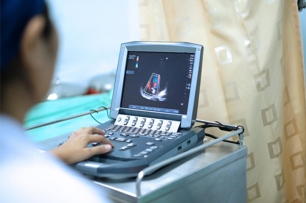
- Directional blood flow
Transesophageal Echocardiography
Transesophageal echocardiography involves passing a slim ultrasound probe down the oesophagus (food pipe). Because the oesophagus lies directly behind the heart, this provides clearer and more detailed images. Transesophageal echocardiography is usually recommended when more detailed images are needed or when Transthoracic echocardiography results are inconclusive.
This modality is commonly used for:
- Detecting blood clots within heart chambers
- Evaluating heart valve infections (endocarditis)
- Assessing prosthetic valves
- Investigating stroke source
- More detailed assessment of structural heart disease
- Real-time guidance during certain heart procedures
Request an Appointment or Make an Enquiry
Whether you’re looking to book an appointment or need more information, we’re here to assist you. Reach out to request an appointment or make an enquiry, and our team will respond promptly to guide you further.